
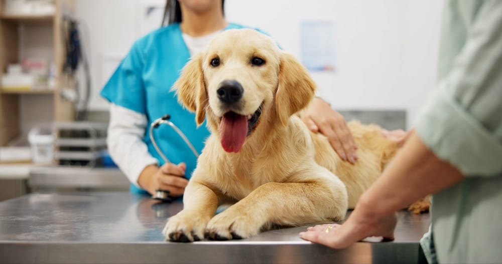
A 90 to 95 percent success rate sounds reassuring, and for good reason. For pets facing a cranial cruciate ligament rupture, TPLO surgery for dogs delivers that outcome in most cases.
But the data tells a clearer story than the statistic alone. What separates successful recoveries from complicated ones is not the surgery itself. It comes down to six specific post-operative actions that dog owners control during the weeks that follow.
Key Takeaways
- TPLO rotates the tibial plateau to neutralize shear force at the stifle joint, without replacing the torn ligament.
- Six post-operative factors, including strict exercise restriction, cold compression therapy, and structured rehabilitation, determine whether bone healing succeeds.
- Roughly 16.5% of patients face complications, but long-term outcomes stay strong across all age groups with proper rehabilitation.
- The surgery gives your dog a strong statistical advantage, but six post-operative actions determine whether that advantage holds.
- Find answers to the most common questions about recovery timelines, candidacy, and post-op care.
How Cranial Cruciate Ligament Rupture Damages the Dog Knee and What TPLO Actually Does {#how-cranial-cruciate-ligament-rupture-damages-the-dog-knee-and-what-tplo-actually-does}
Cranial cruciate ligament disease is a leading cause of pelvic limb lameness in dogs. The stifle joint, or the dog knee, is where the damage occurs. When the cranial cruciate ligament ruptures, it removes the main restraint against cranial tibial thrust.
That thrust is the forward shear force that pushes the tibia out from under the femur with every step. Without it held in check, femoral-tibial translation happens each time the dog bears weight. This leads to pain, cartilage damage, and joint instability that worsens over time.
CCL tears affect many breeds, but Labrador retrievers are among the most common. Unlike an ACL injury in humans, cruciate ligament rupture in dogs often develops through gradual tissue breakdown. A single traumatic event is less common. Many dogs carry a partial tear long before anyone identifies a full rupture.
What the TPLO Surgical Procedure Actually Changes
Tibial Plateau Leveling Osteotomy addresses the problem at its mechanical source. Rather than replacing the torn ligament, the surgical procedure changes the geometry of the tibial plateau itself. A curved cut is made through the proximal tibia. The surgeon rotates the plateau until its angle no longer allows cranial tibial thrust during normal movement.
Once that rotation is complete, a bone plate with locking screws holds the osteotomy site stable. The tibia heals in its corrected position over the following weeks. The result is a stifle joint that stays stable during walking, running, or climbing without the torn ligament.
This is what sets the procedure apart from older approaches like extracapsular repair and lateral suture techniques. Those methods add an external structure to compensate for the torn ligament.
The Slocum model behind this osteotomy changes the joint’s physics so the absent ligament is no longer needed. The caudal cruciate ligament stays intact and functions normally. Joint shear force drops to a level the stifle can handle on its own.
The 90 to 95 percent success rate reflects how well this correction holds. But proper surgical technique is only part of the equation. The rest depends on what happens in the weeks after.
The Recovery Process: Six Post-Operative Factors That Determine Whether Bone Healing Succeeds
As reported in Veterinary Surgery, the recovery process determines most of the outcome. Six post-operative factors sit within your control as a dog owner. Each one directly affects whether your dog reaches full recovery or encounters a complication that sets the timeline back.
Factor 1: Strict Exercise Restriction for the First 8 Weeks
The metal plate and locking screws hold the tibia while bone healing occurs at the osteotomy site. Running, jumping, or stair climbing during this window can disrupt consolidation at the surgical plate. Strict exercise restriction for the first 8 weeks protects the repair.
Short, controlled leash walks are typically allowed from the start. Everything else stops until imaging confirms enough healing to handle increased load.
Factor 2: Elizabethan Collar Use
An Elizabethan collar prevents your dog from licking or chewing the incision. Incision interference is a direct route to surgical site infection, and infection near a bone plate is a serious problem. The collar stays on until the incision fully heals, no matter how much your dog protests.
Factor 3: Cold Compression Therapy
Swelling around the stifle commonly occurs in the early post-operative period. Cold compression therapy applied during the first few days reduces swelling and helps with pain. Most practices give specific timing and duration instructions. Following those directions makes a measurable difference in your dog’s comfort during early recovery.
Factor 4: Structured Post-Operative Physical Rehabilitation
This factor has the strongest research support. One clinical trial showed that structured rehabilitation plus a joint health supplement cut recovery time from 24 to 12 weeks. Rehabilitation includes range-of-motion work, controlled walking, and rehabilitative exercises introduced as bone healing advances.
Veterinary physiotherapy referrals help confirm you perform exercises correctly and progress at the right pace. Skipping this phase is one of the most common reasons dogs take longer to regain normal limb function.
Factor 5: Follow-Up Radiographic Imaging at 8 Weeks
Radiographic images at the 8-week mark confirm whether the osteotomy site has healed enough for more activity. This imaging also lets the veterinary surgeon check for early implant complications. Radiographic healing confirmation is the checkpoint that decides the next recovery phase.
Factor 6: Active Monitoring and Prompt Reporting
Watching your dog’s incision, gait, and comfort after surgery is critical. Contacting your veterinary team immediately when something changes is not optional.
Early reporting of swelling, lameness, or wound changes helps catch complications before they affect the long-term outcome. The six-factor recovery plan only works when your team identifies problems early.
Long-Term Outcomes, Meniscal Injury, and the Complications Dog Owners Must Watch For
Research published in Frontiers in Veterinary Science found that post-operative complications occur in about 16.5% of patients. That figure does not mean the surgery failed. But a meaningful number of dogs will face a complication that needs attention. Owners who spot the signs early consistently have better outcomes than those who wait.
Surgical Site Infection
Surgical site infection is one of the most common major complications. It can develop despite postoperative antibiotics and proper wound dressings when incision care is inconsistent.
Watch for increased redness, discharge, warmth, or swelling at the incision site. Any of those signs call for a same-day contact with your veterinary team. Catching infection early makes a significant difference in recovery.
Tibial Tuberosity Fracture
The tibial tuberosity sits at the front of the tibia, just below the stifle. While the osteotomy consolidates, this area carries increased stress. A tibial tuberosity fracture shows up as a sudden increase in lameness, often without any obvious injury event. This is a key reason strict exercise restriction during the first 8 weeks is non-negotiable.
Meniscal Injury
The medial meniscus sits between the femur and tibia, absorbing load during movement. When the cranial cruciate ligament ruptures, the medial meniscus faces abnormal shear force and can tear. The surgeon treats a tear found during surgery right away. If it develops later, it typically shows as a clicking sound with increased lameness.
Meniscal injury is worth understanding before surgery, not just after. Dogs with cranial cruciate ligament disease often already have meniscal damage. The surgeon will assess and address it during the procedure. Knowing this ahead of time helps set realistic expectations.
Implant Breakage
Implant breakage happens most often when healing stalls and the bone plate takes repeated load without enough support. Confirming radiographic healing at 8 weeks before increasing activity is the primary way to lower this risk.
Long-Term Outcomes Across Age Groups
Gait analysis and ground reaction forces are the standard tools for measuring long-term outcome. Subjective gait analysis combined with force plate data gives a full picture of limb function. A study in PLOS ONE noted that long-term outcomes are comparable across all age groups, including older dogs, when owners follow rehabilitation protocols. Age alone is not a reason to decline the procedure for an otherwise healthy candidate.
Muscle mass loss during recovery is a secondary concern, particularly in older dogs. Structured post-operative physical rehabilitation addresses this directly by rebuilding strength in a controlled, progressive way.
Six Decisions That Make or Break TPLO Surgery for Dogs
TPLO surgery for dogs carries a 90 to 95 percent success rate. That rate is within reach for your dog. The operation corrects the mechanical problem at the stifle joint by eliminating cranial tibial thrust. But the six factors covered here carry that correction through to full recovery.
Exercise restriction protects the bone plate while healing occurs. The Elizabethan collar guards the incision. Cold compression therapy controls early swelling.
Structured rehabilitation cuts recovery time in half. Radiographic imaging at 8 weeks confirms healing before activity increases. And active monitoring catches complications before they change the outcome.
Each factor plays a specific role. Skipping even one creates risk that the surgery alone cannot compensate for.
If your dog has a cranial cruciate ligament rupture, talk to a veterinary surgeon. Schedule a consultation with our team to confirm candidacy and build a recovery plan around your pet.
FAQs
What Is TPLO surgery for dogs?
TPLO stands for Tibial Plateau Leveling Osteotomy. It is a surgical procedure that corrects cranial cruciate ligament rupture by cutting and rotating the tibial plateau, eliminating the shear force that destabilizes the knee joint. The result is a stifle that stays stable without relying on the torn ligament.
How much is TPLO surgery for dogs?
Cost typically ranges from $3,500 to $6,500 per leg, depending on your location, the surgical facility, and your dog’s size. That figure generally includes anesthesia, the procedure itself, and initial follow-up imaging. Pet insurance, payment plans, and financing options can help manage the expense if cost is a concern.
How long does TPLO surgery take for dogs?
The procedure itself typically takes 90 minutes to two hours. Total time at the facility runs longer once you factor in pre-surgical preparation and anesthesia recovery. Most dogs go home the same day or after an overnight observation stay, depending on how smoothly they come out of anesthesia.
How to care for a dog after TPLO surgery?
Strictly restrict activity for the first 8 weeks. Keep the Elizabethan collar on to protect the incision. Apply cold compression therapy as directed to manage swelling. Attend all follow-up imaging appointments. Start structured rehabilitation exercises as your veterinary team introduces them. Monitor the incision daily and report any changes immediately.
Is TPLO surgery worth it for older dogs?
Yes, for most older dogs it is. Research confirms that long-term outcomes after TPLO are comparable across all age groups when rehabilitation is followed. Age alone does not disqualify a dog from surgery. Our veterinary surgeon will evaluate your dog’s overall health to confirm that they are a good candidate.